

Description
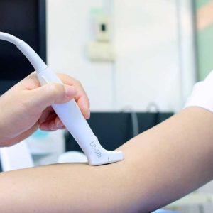
Quick, safe, and pain-free private Knee Ultrasound to assess the Joints, Tendons, Muscles, Bursa, Pain & Range of Movement.
Scans Performed by Specialist Doctors feel confident in the care you receive and gain peace of mind with Same-Day Results & Tailored Advice.
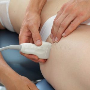
Ultrasonography is a time-efficient and safe method for assessing knee abnormalities.
- It will detect a joint effusion. Evaluation begins with probe placement just cephalad to the patella, allowing identification of the quadriceps tendon in the near field and cortex of the femur in the far-field; between these two structures lies the suprapatellar recess of the knee joint, intercalating between the suprapatellar and pre femoral fat pads. An effusion will cause distension of the visualised recesses with fluid, which may be anechoic or of mixed echogenicity.
- Tendon ruptures. Ruptures of the patellar and/or quadriceps tendon are rare injuries requiring immediate repair to re-establish knee extensor continuity and to allow early motion. Ultrasound is extensively used as a diagnostic tool before surgery on acute traumatic tears of the patellar tendon and quadriceps tendons.
- Meniscal Tear. Ultrasound can also visualise meniscal cysts which can occur secondary to a meniscal tear. Meniscal cysts are small fluid-filled pockets that arise from the meniscus.
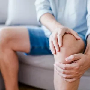
- Osteoarthritis, Ultrasound has been evaluated for its utility in painful episodes in osteoarthritis. Ultrasound has an important role in the evaluation of mechanical arthritis.
- Gout. Ultrasound findings can strongly and independently support the diagnosis of gout and may be useful in the early detection and monitoring of treatments.
- Ganglion cysts about the knee are frequent findings but may be incidental. They are mucin-filled cystic structures that may be unilocular or septate and appear anechoic to hypoechoic. Ultrasound is useful for guidance of aspiration for symptomatic cysts.
- Baker’s cyst. Ultrasound is highly sensitive for the detection of Baker cysts, which are seen as thin-walled fluid collections, sometimes containing debris. The fluid may be anechoic, hypoechoic, or of mixed echogenicity relative to skeletal muscle.
Baker cyst typically communicates with the knee joint, extending between the semimembranosus and medial gastrocnemius tendons. Demonstrating this connection is diagnostic for a Baker cyst with 100% accuracy and can eliminate false-positive diagnoses and misinterpretation of other cystic structures or masses as Baker cysts. A leaking Baker cyst shows fluid tracking between the muscles and along the subcutaneous tissues.
- Muscles. Ultrasound is useful for the evaluation of muscle injury, including demonstration of partial- and full-thickness tears of the muscle and tendon. A partial tear will appear as an anechoic cleft or hypoechoic collection in the belly of the muscle. Ultrasound can be used to evaluate for herniation of muscle through the fascia, a finding that is sometimes transient and therefore particularly amenable to real-time sonographic evaluation during which muscle flexion can evoke the abnormality. Ultrasound is useful in the evaluation and follow up of partial- and full-thickness tears of the medial head of the gastrocnemius ‘‘tennis leg’’, showing disruption at the origin and commonly associated with a hematoma or fluid collection between the gastrocnemius and the soleus muscles.