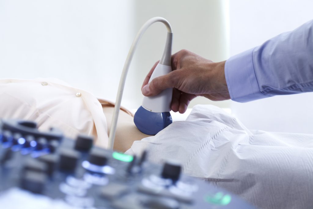
Testes/scrotum ultrasound is a non-invasive diagnostic technique that involves sound waves to examine the testicles and scrotum. It plays an important role in determining various conditions affecting the male reproductive system. This article explores how Testes/Scrotum Ultrasounds are performed for diagnoses.
What Conditions or Diseases a Testes/Scrotum Ultrasound Can Determine
This scan helps in diagnosing specific conditions, including testicular torsion, testicular tumors, epididymitis, varicocele, and hydrocele. Let’s discuss these conditions in detail.
Testicular Torsion
Testicular torsion occurs when the spermatic cord twists, leading to a disruption in blood flow to the testicle. Testes/scrotum ultrasound is instrumental in diagnosing this condition by evaluating the blood flow to the affected testicle. Using Doppler ultrasound, the radiologist can identify the absence or reduced blood flow to the twisted testicle, aiding in prompt diagnosis and intervention.
Testicular Tumors
Testes/scrotum ultrasound is a valuable tool for detecting and characterizing testicular tumors. The ultrasound images can reveal the presence of solid masses or cystic lesions within the testicles. By analyzing the size, shape, and vascularity of the mass, the radiologist can determine if it is likely benign or malignant. Detailed evaluation, such as testicular biopsy or tumor marker testing, may be necessary for definitive diagnosis.
Epididymitis
Epididymitis refers to the inflammation of the epididymis, a coiled tube located behind the testicle. Testes/scrotum ultrasound helps in diagnosing epididymitis by detecting enlargement and increased blood flow to the affected epididymis. It can also assess for the presence of abscesses or fluid collections within the epididymal region, aiding in appropriate treatment planning.
Varicocele
Varicoceles are dilated veins in the scrotum that can affect male fertility. Testes/scrotum ultrasound plays a crucial role in identifying and evaluating varicoceles. The ultrasound images show the enlarged veins and their location within the scrotum, according to PubMed. By assessing the severity of the varicocele and its impact on blood flow, the radiologist can provide valuable information for fertility evaluations and potential treatment options.
Hydrocele
Hydrocele refers to the accumulation of fluid around the testicle, leading to scrotal swelling. Testes/scrotum ultrasound helps in determining the presence and characteristics of hydroceles. The ultrasound images show the fluid-filled sac surrounding the testicle and aid in differentiating hydroceles from other scrotal masses. The size and extent of the hydrocele can be assessed, assisting in treatment decisions, including potential surgical intervention.
Summary
Testes/scrotum ultrasound is a versatile imaging technique that enables the diagnosis and evaluation of various conditions affecting the male reproductive system. By utilizing sound waves and advanced imaging technology, this non-invasive procedure provides detailed images of the testicles and scrotum.
The evaluations from testes/scrotum ultrasound aid healthcare professionals in accurate diagnosis, treatment planning, and fertility evaluations. With its effectiveness and safety, testes/scrotum ultrasound remains an invaluable tool in the field of male reproductive health. Contact Us if you want answers to your queries and get our help.


