DVT is a deep vein thrombosis (DVT) which is a blood clot formation in the deep veins and can occur in any area of your body, especially your legs. When a blood clot restricts the blood flow, it is known as a thrombus. Blood clots that form in a deep vein are known as venous thrombosis. But it is more likely to develop in deep leg veins that run through the muscles of the calf and thigh. Your doctor may ask for a DVT ultrasound.
Let’s explore more.
What are the Causes of Deep Vein Thrombosis
The following reasons can cause the Deep Vein Thrombosis
- Family history of blood clots formation
- Being overweight or obesity
- A Polycythemia Vera that develops bone marrow for making many blood cells
- Suffering a long-time catheter, a tube in blood vessels
- Sitting for a long period, for example, travel
- A deficiency of vitamin C increases the risk of a blood clot
- An injury to the vein
- Having a surgery
- An inflammation of the walls of the vein
How to Diagnose the Deep Vein Thrombosis?
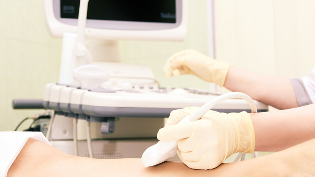
Based on your condition and the symptoms you experience, your doctor may suggest a DVT Ultrasound to diagnose the causing factor. An ultrasound involves sound waves that give an image of the blood flow through your veins and arteries. It is not a harmful diagnostic procedure. Do you know why? Well, it does not use radiation.
Duplex ultrasonography helps to detect blood clots in the deep veins and also provides real-time video or images. Your healthcare provider may ask you to wear a hospital gown. There is only your leg that needs to be exposed for evaluation.
It allows your healthcare provider to move the wand on the surface of the legs. Behind your knee, there is more possibility of DVT. A gel is used to apply on the skin surface that allows the probe to move without the formation of bubbles. The probe is moved back and forth direction to check the blood clot in deep veins as sound waves produce images on the computer screen.
You may need to change your body positions during DVT ultrasound as it helps to view the area and make it easy to detect blood clot formation.
An ultrasound may take less than 30 minutes to identify a deep vein thrombosis. If your doctor suspects a moving or growing thrombus, you may need more tests to find out the thrombus. Do you know why?
The moving thrombus is dangerous as it can pass to the heart and can cause serious health risks. Having more tests can help identify deep vein thrombosis in different parts of your body and also assist in its removal. If you experience DVT symptoms, ensure you get an ultrasound, or an untreated condition cause serious health complications, according to PubMed.
Your doctor may ask for other tests, such as:
Venography – A dye injected into the vein that helps to detect the blood flow and locate blood clots.
MRI – It involves radio waves and frequencies to get detailed images of the body area.
CT Scan – It uses computer equipment and X-rays to get three-dimensional images.
Pulmonary Ventilation Scan – A two-series lung test to check the blood flow and how it functions.
What is the Accuracy Rate of DVT Ultrasound?
In Above the knees, ultrasounds can give the best results in identifying blood clots. The accuracy rate is high when it comes to finding the blood clot in the above area of the knees. Once your doctor detects the clot, you’ll be given a treatment plan. But there is only a 60% chance of locating a blood clot in the leg calf as it is difficult to find out in such areas.
What are the Symptoms of Deep Vein Thrombosis DVT?
Some symptoms may indicate that there is a formation of a blood clot in your deep veins, such as:
- Swelling on your leg (calf or behind your knee)
- Warm feeling around the area of the clot
- Redness on some specific area of your leg
- Having pain or tenderness in the clot location
When you notice some changes in your leg, ensure you get yourself checked. Do you know why it is important?
Some blood clots may not move once they form in your legs, and they can be treated. But some blood clots are moving and can go in your heart direction and then to your lungs. It may cause serious health issues and put you at high risk of pulmonary embolism (PE).
The Bottom Line
A Deep Vein Thrombosis DVT ultrasound can help to detect blood clots by 90% in the above area of your knees but only 60% in your calf. Your healthcare provider can ask for other tests if there is no blood clot detected or suspected to be moving. If you experience the above symptoms, visit your healthcare provider as early treatment can reduce the pain and swelling. At RIZ Ultrasound, you can get the best ultrasound services in Glasgow. You can Contact Us if you need any help.