A neck ultrasound is an imaging test that helps to view the internal structure. It uses sound waves that create images of organs inside the neck. The neck area includes organs, such as the thyroid gland, deep neck lymph nodes, soft tissue organization of the neck jugular arteriovenous system, and salivary gland.
Healthcare providers order a Neck Ultrasound if they experience any symptoms or signs related to organs inside your neck. A transducer is used that emits sound waves. These waves go inside the neck and do not cause any harmful effects.
This article will help you learn more about it.
Why a Neck Ultrasound is Done?
A neck ultrasound is beneficial when it comes to diagnosing a number of diseases or abnormalities in the neck. An abnormal function of the thyroid or an inflamed thyroid can cause many symptoms and signs. A neck ultrasound helps to detect the size and appearance of thyroid nodules, determine lesions in the thyroid gland, and evaluate if there is any other medical condition affecting the thyroid.
Lymph nodes play important roles but they can get affected by a disease. Sometimes, an examination of the lymph nodes helps to diagnose the cause. Performing an ultrasound can help to view the size. It also helps to observe the wall of the esophagus in the neck and assess the jugular arteriovenous systems that can be caused by thrombosis.
An ultrasound is also useful to diagnose nasopharyngeal cancer by viewing the changes in the abnormalities of the cervical lymph nodes, according to NCBI.
When there is a change in the size, sensation, and color of neck lymph, it indicates a disease associated with neck organs. Enlarged lymph often indicates thyroid cancer, goiter, benign thyroid, and autoimmune diseases.
Healthcare providers suspect the condition by the symptoms and signs but order a neck ultrasound to diagnose the causing factor.
How to Perform a Neck Ultrasound?
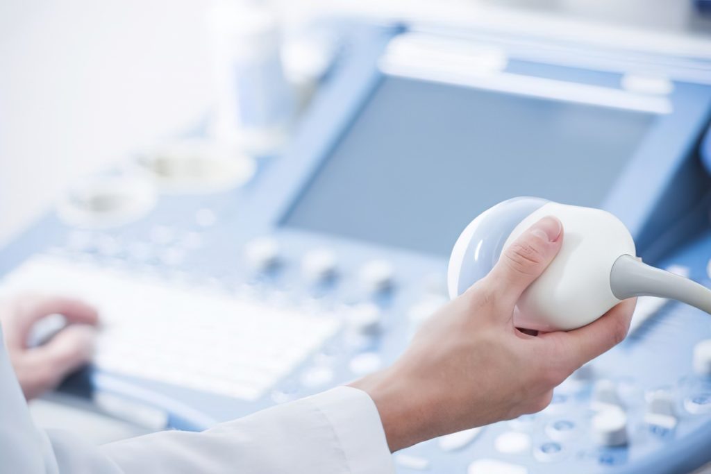
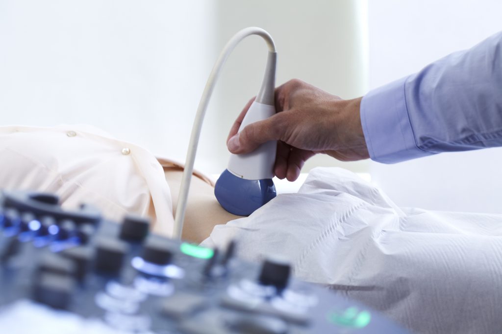
Neck ultrasound is performed using a device, called a transducer. It uses high-frequency sound waves that penetrate your neck and reflect when they are hit by a dense object. A technician uses a gel to apply on your neck. It helps a transducer to move in and forth direction. Using a gel eliminates the risk of air gaps between the transducer and your skin which causes blurred images.
A professional will ask you to lie down, facing head to the roof. This position helps a technician to move the transducer back and forth direction to capture every angle of the neck area.
It takes 20 to 30 minutes to perform and get detailed images.
How to Prepare for a Neck Ultrasound?
An ultrasound is a pain-free and non-invasive procedure that does not require any special preparations. Unlike other blood sugar tests, you do not need to fast before a neck ultrasound. But you need to avoid some of the foods that can cause damage to your thyroid. Eating too hold or cold foods can cause harm to your thyroid. Ensure that you prefer a healthy diet and avoid eating a fermented or high-salt diet that is not a good choice for the thyroid before having an ultrasound.
Experts at Private Neck Ultrasound Scan in Glasgow share that an ultrasound of your neck can also be performed with other diagnostic procedures.
Takeaway
Neck ultrasound is a modern yet the best diagnostic technique that helps to detect diseases or conditions associated with internal neck organs. If you experience symptoms like swollen neck area or pain while swallowing food, ensure you get medical help. An early evaluation can help to get the treatment at the right time and be more effective when it comes to results.


