The human hand consists of dozens of tendons that connect muscles to bones and make hand movements possible. Tendons are connective tissues that may rupture due to an injury, resulting in pain and swelling in the hand. Tendon injuries limit the motion of the hand and reduce grip while holding objects. A hand and wrist ultrasound can help to diagnose potential injuries. The article will describe common types of tendon injuries and the role of ultrasound in diagnosing them.
What are Tendons?
Tendons are connective tissues that connect muscles to bones in the body. Muscle contraction causes the relevant tendon to pull the bone and, thus, brings about movement. Two types of tendons are involved in the mobility of hands:
Flexor tendons: The flexor tendon moves from the forearm across the palm through the wrist. The flexor tendon helps in bending the fingers.
Extensor tendons: The extensor tendon moves from the forearm across the back of the hand into the thumb and fingers. It helps straighten the thumb and fingers.
There are nine flexor and twelve extensor tendons in the human hand. Any damage to these tendons may cut or rupture the tendon, leading to loss of movement.
Common Hand Tendon Injuries
You might get a tendon injury due to trauma, handling sharp objects, or sports. Following are some common injuries that affect hand joints:
Mallet Finger
Mallet finger results from a ruptured extensor tendon at the terminal phalanx. This condition results from overbending of the distal interphalangeal joint. A bowed fingertip and pain in the back of distal interphalangeal joints characterise Mallet’s finger.
Boutonniere Deformity
Boutonniere deformity is when the joint adjacent to the knuckle is bent inwards, and others are bent outwards. This type of injury occurs due to a rupture of the extensor muscle at the middle phalanx. Boutonniere deformity results from forced bending of the proximal interphalangeal joint against the extension, causing the central slip of the tendon to rupture. Individuals with this deformation cannot extend the middle joint of the finger.
Jersey Finger
Jersey finger results from a traumatic tearing of the digitorium profundus muscle from the terminal phalanx. Patients with this type of injury feel pain while bending the distal interphalangeal joint and inability to stabilise the proximal interphalangeal joint. The Jersey finger is named so because it occurs when a player seizes the jersey of another player with bent fingers.
Gamekeeper’s Thumb
The gamekeeper’s thumb is a hyperabduction injury to the ulnar collateral ligament of the thumb knuckles. The injury is associated with extreme abduction stress to the metacarpophalangeal joint of the thumb and overextension. An individual with this deformity presents pain in the ulnar region and reduced ability to pinch.
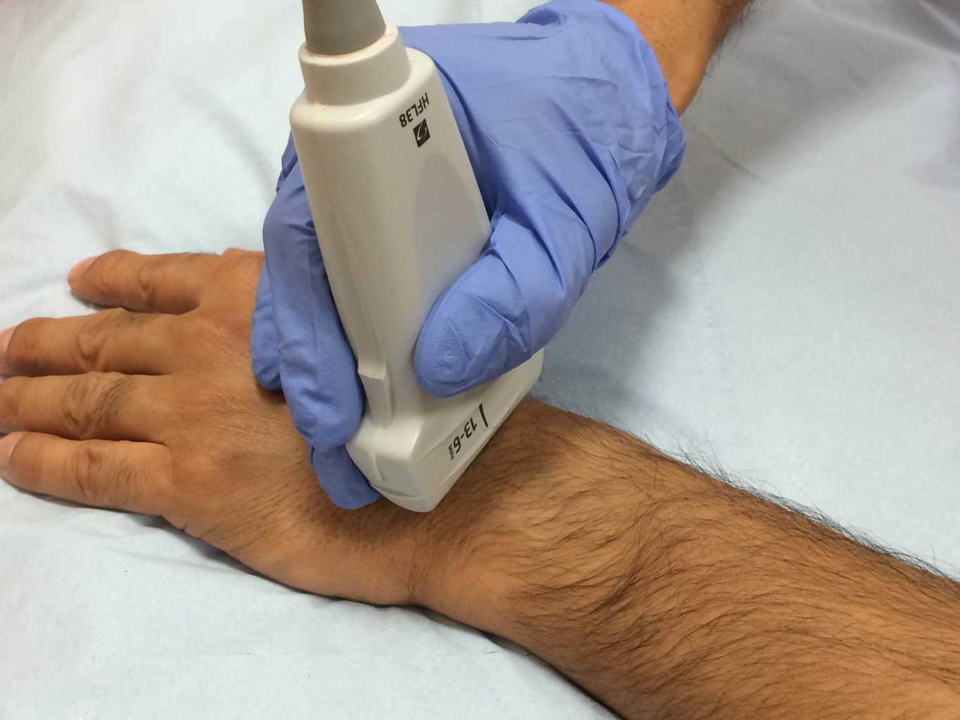
Role of Ultrasound in Detecting Hand Tendon Injuries
Despite various imaging techniques, High-resolution ultrasound has emerged as a modern diagnostic tool to detect hand tendon injuries. Hand and wrist ultrasound is a non-invasive and low-cost technique that provides good resolution and image quality. Preoperative evaluation of tendon injury through hand and wrist ultrasound is essential for identifying the tendon injury type and planning reconstructive surgery. Moreover, post-operative ultrasound helps detect the solidarity of the repaired tendons.
Final Thought
Hand tendon injuries are common in athletes and sportsmen. Though they seem minor, these tendon injuries cause pain and limitations in the extent of movement. High-resolution ultrasound assessment provides subtle details about the type of tendon injury. The technique also helps in the decision of surgery and evaluation of tendon repair after surgery.